
Fig. 2. PTEN sensitizes cells to TNF-induced apoptosis.
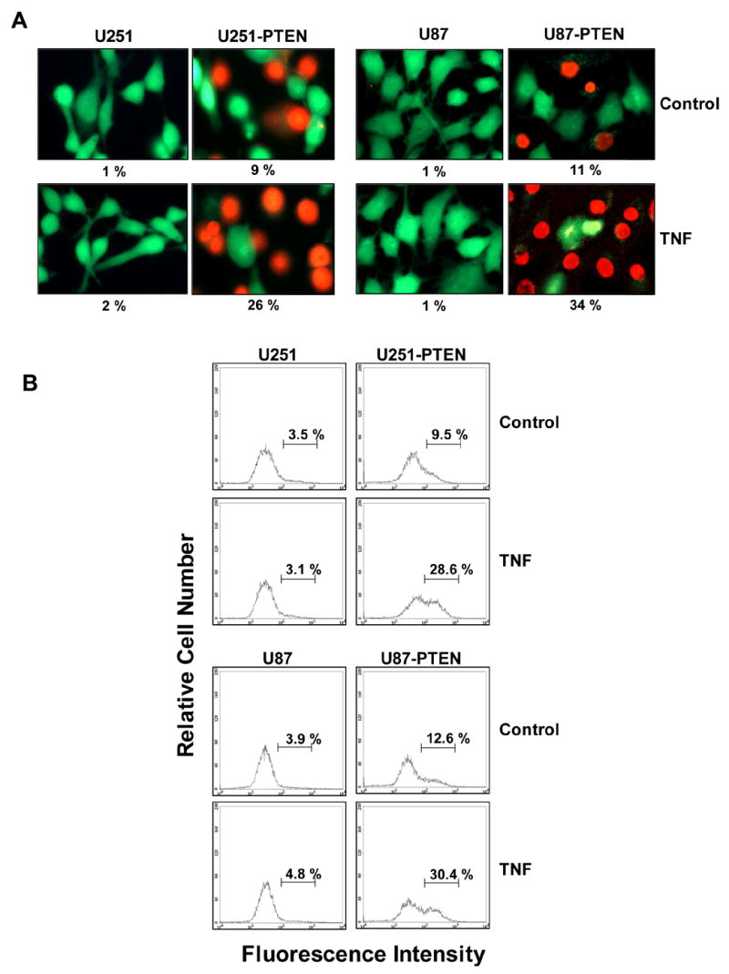
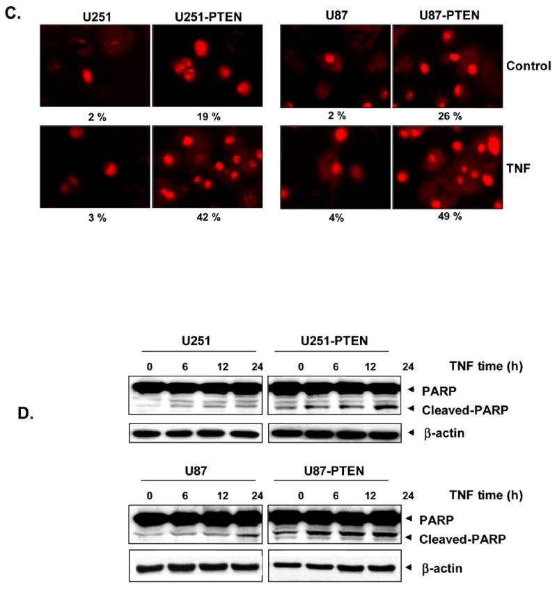
A, U251/U251-PTEN and U87/U87-PTEN cells (1 x 10⁵ cells/ml) were treated with 1 nM TNF for 16 h. Cells were stained with the Live/Dead reagent for 30 min. Cells were analyzed under a fluorescence microscope. B, U251/U251-PTEN and U87/U87-PTEN cells were treated with 1 nM TNF for 16 h and then underwent annexin V staining. Cells were washed, incubated with FITC-conjugated anti-annexin V antibody, and then analyzed by flow cytometry. C, U251/U251-PTEN and, U87/U87-PTEN cells (1 x 10⁵ cells/well) were treated with 1 nM TNF for 16 h. Cells were washed with phosphate-buffered saline, air-dried, fixed, permeabilized, and then stained with TUNEL assay reagent, after which they were analyzed under a fluorescence microscope. D, U251/U251-PTEN and, U87/U87-PTEN cells (5 x 10⁵ cells/well) were treated with 1 nM TNF for the times indicated, and whole-cell lysates were subjected to SDS-PAGE. Western blot analysis was performed using anti-PARP antibody. β-actin was blotted as a loading control.