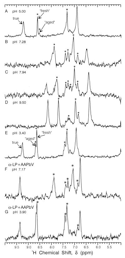
Figure 2.
1D 1H/13C HMQC NMR spectra vs. pH of partially denatured ≈1 mM {13C, 15N} His-labeled α-lytic protease at 25°C. (F and G) Sample after inhibition with ≈2-fold excess of MeOSuc-Ala-Ala-Pro-boroVal (AAPbV). The asterisks denote extraneous peaks because of denatured or inactive enzyme. This sample, which had been frozen for 3 yr, remained at each pH value for 24 h or longer.

