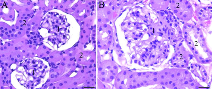
Figure 3.
Photomicrograph of kidney sections of female mice. (A) Wild-type mouse. (B) B6.129S6- Naglutm1Efn/J mutant mouse. Note the large, distended glomerulus (1) with increased mesangium as well as the cells containing vacuolar or granular cytoplasm within the glomerular epithelium and adjacent renal tubules (2) in (B) compared with the normal-appearing renal tissue of the wildtype mouse (A). Hematoxylin and eosin staining; scale bar, 50 μm.

