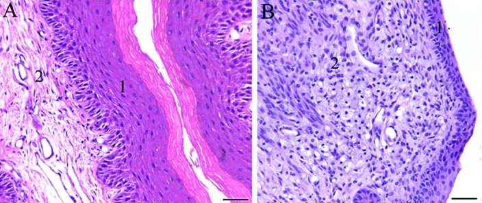
Figure 5.
Proximal urethral–vaginal submucosa. (A) Wildtype mouse. (B) B6.129S6- Naglutm1Efn/J mutant mouse. 1, Squamous epithelium of the vagina; 2, submucosa. Note the abundant amount of submucosal mononuclear infiltrate composed of large foamy granular cells which attenuate the squamous mucosa in (B) compared with the normal histology in (A). Hematoxylin and eosin staining; scale bar, 100 μm.

