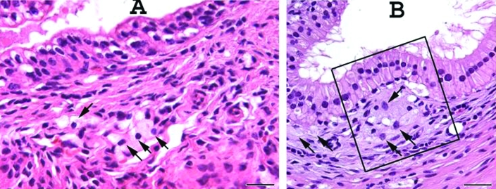
Figure 6.
Urogenital tract structures in prostatic region of a B6.129S6- Naglutm1Efn/J mutant mouse. (A) Subtle mononuclear cell infiltrate (arrows) in the lamina propria of an intraprostatic duct. (B) Subepithelial accumulation of mononuclear cell infiltrate (arrows), which elevates the epithelium toward the lumen of the ductus deferens (boxed area). Note that the infiltrate within the lamina propria is less prominent than that in the urogenital structures of female mutant mice (Figure 5). Male mice had only single granular cells or small clusters of granular cells compared with the abundant infiltrate in female mice. Scale bar, 25 µm.

