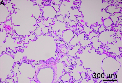
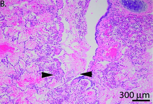
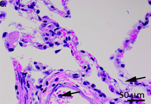
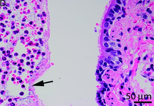
Figure 1.
(A and C) Acute fibrinous interstitial pneumonia with bacteria in major pulmonary vessels and septal capillaries, consistent with hematogenous seeding of lung. (B and D) Acute suppurative bronchopneumonia consistent with primary pneumonia rather than hematogenous seeding of lung. Arrows point to bacteria, and arrowheads point to fibrin and neutrophils in a bronchiole; this animal also had slight alveolar hemorrhage. Magnification, ×40 (A and B), ×100 (C and D).


