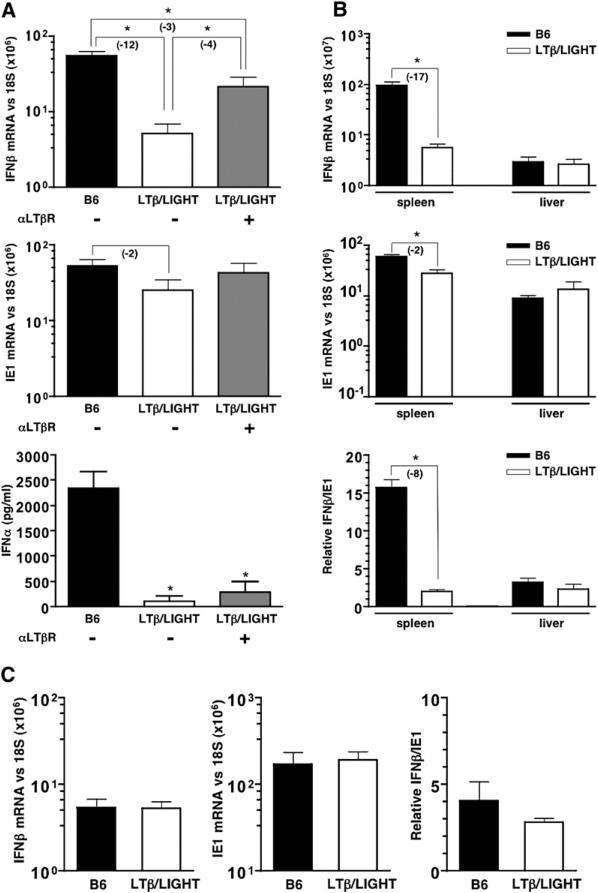
Figure 2. LTβ/LIGHT−/− Mice Are Defective in Their Initial IFNαβ Response to MCMV.
Wild-type (B6) and LTβ/LIGHT−/−-deficient mice were infected with MCMV plus or minus treatment with an agonistic anti-LTβR antibody (±αLTβR) at the time of infection. Spleens were harvested 8 hr postinfection and (A) IFNβ and ie1 mRNA levels were determined by qPCR. Serum INFα levels measured by ELISA (n = 4−6 mice per group from two independent experiments, mean ± SEM) (bottom panel). (B) Spleens and livers from MCMV infected B6 and LTβ/LIGHT−/− mice were analyzed by qPCR 8 hr postinfection for IFNβ and IE1 mRNA levels (top two panels). The ratio of IFNβ to ie1 mRNA (×100) is shown (bottom panel, n = 3 mice per group, mean ± SEM). (C) Spleens from B6 and LTβ/LIGHT−/− mice were harvested 48 hr postinfection with MCMV, and IFNβ and ie1 mRNA levels were determined by qPCR (n = 3 mice per group ± SEM). Mice were injected with 2 × 10⁵ PFU of MCMV i.p. for all experiments. Statistical significance (*p < 0.05) was determined using the Student's t test, and fold differences are shown in parentheses.

