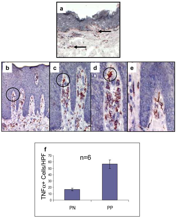
Figure 2. TNFα expression in PN and PP skin.
Cryopreserved sections of PN skin (panel a) and PP skin (panels b–e) were immunostained to detect TNFα. Note the markedly increased number of TNFα + cells in PP skin (b) compared to PN skin (a). The majority of TNFα + cells in PP skin are located in the papillary dermis (b–e). Note that progressively increased magnification is provided in panels b–d using a black circle for orientation. Many of the TNFα + cells had a dendritic morphology (e), whereas others had a plump macrophage-like appearance (d). No epidermal keratinocyte immunoreactivity for TNFα was observed.
Quantitation of TNFα immunoreactive cells involving six different subjects (n=6) for PN skin (17±2 cells/HPF) and PP skin (57±7 cells/HPF) revealed significant differences between PN and PP skin (p<0.05) as depicted in (f).
These results indicate PP skin is characterized by an increased density of TNFα + mononuclear cells that infiltrate the papillary dermis and focally extend into the epidermal compartment.

