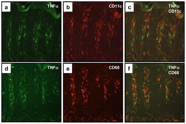
Figure 3. Co-localization of TNFα expressing cells within CD11c+ dendritic cells and CD68+ macrophages.
Cryopreserved sections of PP skin were subjected to two-color immunofluorescence staining in which TNFα expressing cells were labeled green (FITC; a, d), and the CD11c+ (b) or CD68+ (e) mononuclear cells labeled red (rhodamine). Note the similar distribution patterns and morphological profiles, as well as dual fluorescence imaging that highlight the co-expression of TNFα in both CD11c+ (c) as well as CD68+ (f) cell subsets.

