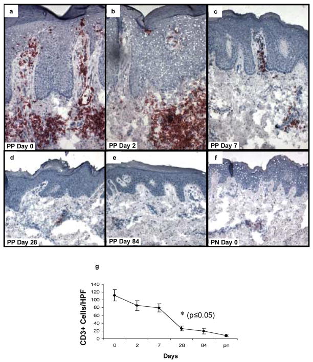
Figure 6. Expression and kinetic profile highlighting temporal reductions in density of CD3+ T cells in psoriatic patients before and after treatment with adalimumab.
Untreated PP skin is characterized by influx of CD3+ T cells, located both in the upper dermis with focal extension into the epidermis. Following treatment with adalimumab, the number of T cells begins to decline on days 2 and 7 (b, c), with a more pronounced reduction on days 28 and 84 (d, e) where they resemble the density in PN skin (f). The earliest time point with statistically significant reduction in CD3+ cell counts compared to untreated samples is day 28 (g; n=6; asterisk, p<0.05).

