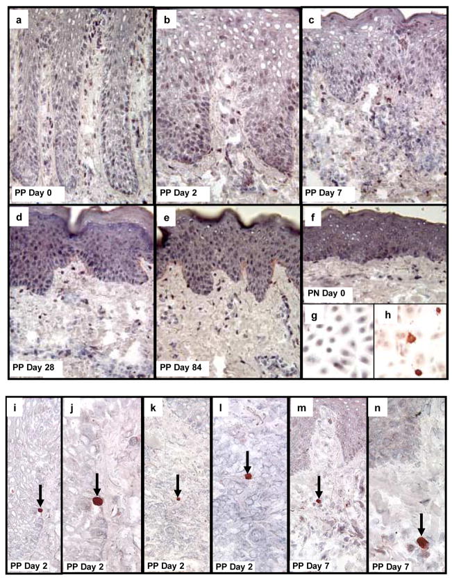
Figure 7. Detection of activated caspase 3 as a marker for cells undergoing apoptosis in psoriatic patients before and after treatment with adalimumab.
Untreated PP and PN skin are characterized by the absence of cells in either the epidermis or dermis undergoing apoptosis because no activated caspase 3 positive cells are present in the tissue samples (a, f). Moreover, following treatment with adalimumab, the reduction in epidermal thickness as well as the decreased density of immunocytes (dendritic cells, macrophages, T cells) was also not accompanied by and prominent increase in detectible cells expressing activated caspase 3 (b–e). As a positive control, cultured human keratinocytes were immunostained before (g), and after, (h) UV-light to confirm detection of activated caspase 3. Note the strong cytoplasmic immunostaining for activated caspase 3 in keratinocytes undergoing apoptosis following UV-light (h).
In skin tissue sections, occasional, rare scattered cells expressing bright red anti-activated caspase 3 reaction product was detected in epidermal keratinocytes (i, j; arrows depict basal layer keratinocyte undergoing apoptosis in PP skin 2 days after adalimumab treatment. A rare dermal lymphocyte expressing activated caspase 3 was also identified at day 2 in treated PP skin (k, l; arrows depict apoptotic lymphocyte). A rare macrophage-like cell expressing activated caspase 3 is shown in (m, n) on day 7 in treated PP skin.

