
Figure 4. Marker Conconversion Analysis of DGR Retrohoming.
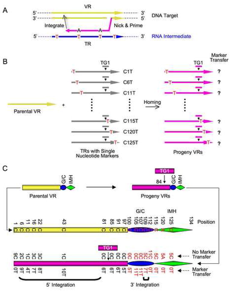
(A) Strategy to identify cDNA integration sites at the 3′ and 5′ ends of VR via marker coconversion. Markers introduced into TR (red Ts) are transferred to VR only if they are located between 3′ cDNA priming and 5′ cDNA integration sites.
(B) Schematic of coconversion experiments. Single C to T markers in donor TRs are indicated. All TRs are tagged with TG1 at position 84.
(C) Homing assays were performed following BPP-1d single-cycle lytic infection of RB50 cells transformed with marked donor plasmids. Results from PCR homing assays are shown in Figure S10 and marker coconversion data are summarized here. Nucleotides at relevant positions in the wild type parental VR are shown in the center. At bottom, the number of progeny VRs with a transferred marker, or no transferred marker at specific sites are shown. Deduced cDNA integration regions at the 3′ and 5′ ends of VR are indicated by brackets.