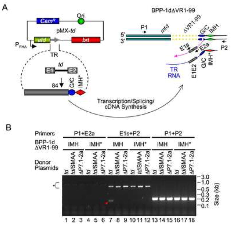
Figure 5. cDNA Integration at the 3′ End of VR.

(A) Experimental design. Donor plasmid pMX-td contains the td group I intron at position 84 in TR, and prophage BPP-1dΔVR1-99 lacks the first 99 bp of VR. cDNA integration at the 3′ or 5′ end of VR was assessed in PCR assays using the same primer pairs described in Figure 1D. (B) VR lacking the first 99 bp supports cDNA integration at the 3′ end, but not the 5′ end. BPP-1dΔVR1-99 (IMH) and BPP-1dΔVR1-99IMH* (IMH*) lysogens transformed with the indicated donor plasmids (described in Figure 1E) were induced with mitomycin C for 2 hrs. Total nucleic acids isolated from induced cultures were assayed for cDNA integration by PCR using primers shown in (A). Pink arrow, cDNA intermediate. *, Brt-independent PCR artifacts.
