
Figure 1.
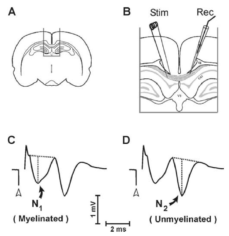
Placement of electrodes for callosal CAP recording, and measurement technique for N1 and N2 CAP components. A., B. Stimulating and recording electrodes were positioned in midline corpus callosum of brain slices separated by approximately 1.0 mm. C. Quantification of the myelinated (N1) wave component. D. Quantification of the unmyelinated (N2) wave component. In (C.) and (D.) CAP amplitude was measured as vertical distance from the local negative peak to a tangent joining preceding and following positivities. Open arrowheads indicate time of stimulus onset.