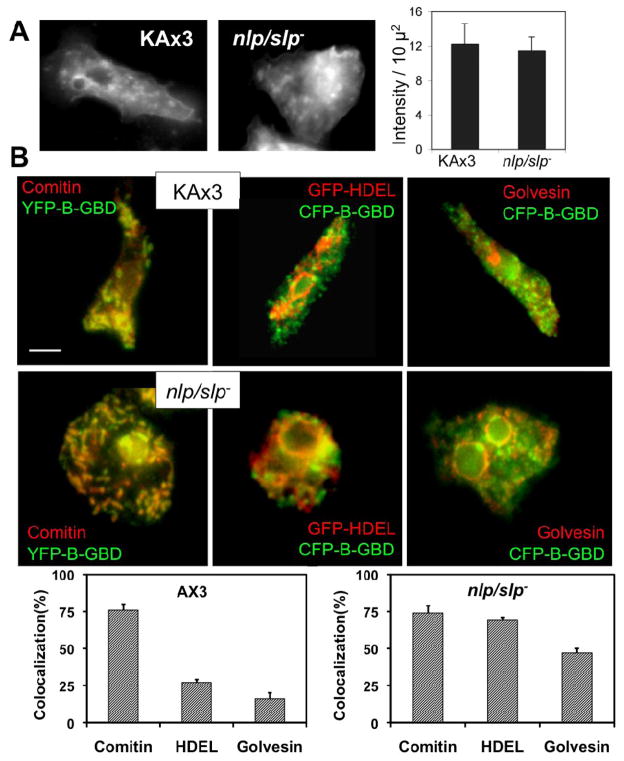
Figure 3.
(A) Membrane endocytosis by aggregation-competent cells determined using FM1-43X. Ax3 or nlp/slp- cells were incubated with FM1-43X for 2 min and washed, followed by fixation for image collection. Cells were fixed after 2 min incubation with FM 1-43 followed by a 10 min delay to allow for endocytosis. Fluorescence intensity of FM1-43X dye in cells was measured and shown in the graph. Error bars represent SEM (n=10). (B) Colocalization of CFP-B-GBD with GFP-HDEL and golvesin-c-GFP in nlp/slp- cells. GFP-HDEL and golvesin-c-GFP overlaps with CFP-B-GBD in nlp/slp- cells, but not in KAx3 cells. At least 15 cells from two independent experiments were analyzed for colocalization as decribed in Methods section. The percent colocalization calculated among areas is shown for the percentage of YFP-B-GBD that colocalized with comitin, GFP-HDEL, or golvesin-c-GFP respectively.

