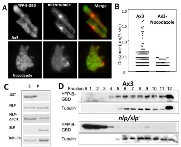
Figure 7.
(A) Distribution of vesicles decorated with YFP-B-GBD in chemotaxing or nocodazole-treated cells. Cells expressing YFP-B-GBD in aggregation stage were fixed and stained with rabbit anti-tubulin antibody. The YFP-B-GBD and microtubule images were superimposed for assessment of localization of YFP-B-GBD vesicles in a close proximity to microtubules. (B) Tracking of YFP-B-GBD vesicle movements in KAx3 or nocodazole-treated cells by live cell imaging. Speed of vesicle movement in wild type or cells treated with nocodazole was measured and plotted. Wild type cells showed brief periods of rapid directed movement leading to high speed (>0.25 μm/sec) that is absent in nocodazole-treated cells. (C) Binding of NLP/SLP and a NLP mutant to microtubule. GST-NLP or SLP were incubated with paclitaxel stabilized-microtubules. Supernatants (s) and pellets (p) were collected after ultracentrifugation over a 15% sucrose cushion and analyzed by SDS-PAGE for presence of GST-NLP or SLP by SDS-PAGE. (D) Cosedimentation of YFP-B-GBD with MT on a sucrose gradient. Lysates of wild type or nlp/slp- cells expressing YFP-B-GBD were applied onto 15–40% sucrose gradient and fractions were collected. YFP-B-GBD in fractions was detected by western blot with anti-GFP antibody.

