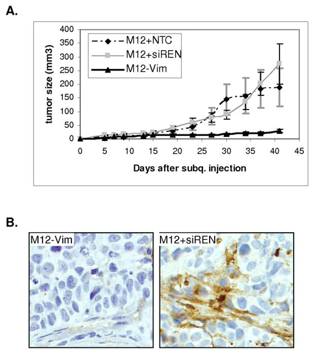
Fig. 4.
Tumorigenic properties of M12-Vim cells are reduced in athymic mice. (A) Tumor formation following subcutaneous injection of 1 × 10⁶ M12-Vim cells (6 mice) was compared to the injection of cells containing vector only M12+siREN (4 mice) or M12+NTC, a non-targeting RNA control, (5 mice). Tumor growth was monitored by caliber measurement each 4 to 5 days for up to 42 days. All animals displayed tumors albeit of varying size and tumor volume (mm3), calculated as described in Materials and Methods. The standard error of the mean is shown as error bars. (B) immunofluoresence staining with human vimentin antibody of paraffin-embedded M12+siREN (left panel) and M12-Vim (right panel) tumors retrieved from nude mice at time of euthanasia (42 days). Magnification is at 400X.

