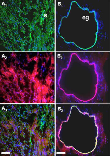
Figure 1.
Simultaneous immunofluorescent staining of COX-2, vimentin, and cytokeratin-8 in ectopic endometrial tissue. COX-2, vimentin, and cytokeratin-8 were immunostained as described in Materials and Methods, and DAPI (blue) was used for counterstaining. Note the green fluorescence corresponding to vimentin (A1) or cytokeratin-8 (B1) and the red fluorescence corresponding to COX-2 (A2 and B2). Superposition of the green and red signals shows simultaneous immunostaining between COX-2 and vimentin (A3) and COX-2 and cytokeratin-8 (B3). Scale bar, 50 μm. eg, Epithelial gland; s, stroma.

