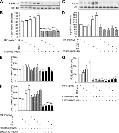
Figure 5.
ERK-1/2 and p38 MAPK phosphorylation in human endometriotic stromal cells in response to MIF, and effect of MAPK inhibition on COX-1 and COX-2 mRNA expression and PGE2 secretion. After starving, cells were preincubated for 45 min with or without PD98059 (50 μm), a specific inhibitor of ERK-1/2 MAPK, or SB203580 (50 μm), a specific inhibitor of p38 MAPK, before MIF addition at a final concentration of 0, 1, 10, 25, or 50 ng/ml. Thirty minutes later, protein extraction and Western blotting were performed as described in Materials and Methods. Representative Western blot of ERK-1/2 MAPK (A) and p38 MAPK (C) are from patient nos. 6 and 3, respectively (Table 1). Densitometric analysis data are from three different endometriotic cell cultures for ERK-1/2 MAPK (nos. 6–8, Table 1) and p38 MAPK (nos. 3, 4, and 10, Table 1). MIF increased ERK-1/2 MAPK (B) and p38 MAPK (D) phosphorylation, and pretreatment with PD98059 or SB203580 completely abolished MIF-induced activation of ERK-1/2 and p38 MAPKs, respectively. *, P < 0.05 vs. control medium. +, P < 0.05; ++, P < 0.01; and +++, P < 0.001 are significantly different from cells incubated with the same concentration of MIF, using ANOVA and the Bonferroni’s multiple comparison test post hoc. To evaluate the effect of MAPK inhibition on COX-1 and COX-2 mRNA expression and PGE2 secretion, endometriotic cells were preincubated for 45 min with or without PD98059 (50 μm) or SB203580 (50 μm) before incubation with MIF (0, 1, 10, or 50 ng/ml) for an additional 12 or 24 h. COX-1 and COX-2 mRNA levels were quantified by quantitative real-time PCR using GAPDH as an internal control, and PGE2 secretion was measured in the culture supernatants using EIA. Data are from three different endometriotic cell cultures (patient nos. 4, 8, and 10, Table 1). Whereas no statistically significant change in COX-1 mRNA expression was noted (E), MIF significantly induced COX-2 mRNA expression, and pretreatment with SB203580 but not PD98059 significantly reversed MIF stimulatory effects (F). G, MIF increased PGE2 secretion, and MIF-induced PGE2 liberation was completely abolished by pretreatment of PD98059 or SB203580. *, P < 0.05; **, P < 0.01; and ***, P < 0.001 are significantly different from the control medium, which was devoid of stimuli (0 ng/ml MIF); +, P < 0.01; ++, P < 0.001 are significantly different from cells incubated with the same concentration of MIF, using ANOVA and the Bonferroni’s multiple comparison test post hoc.

