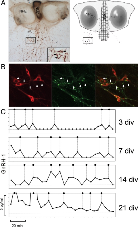
Figure 1.
Kinetics of GnRH-1 secretion. A, GnRH-1 immunostaining of a nasal explant obtained from embryonic d 11.5 mouse, maintained for 7 div, and the corresponding schematic showing nasal pit epithelium (NPE) (ovals) and the nasal midline cartilage (NMC) (meshed area), surrounded by mesenchyme. GnRH-1 neurons (dots) migrate from NPE and follow olfactory axons to the nasal midline cartilage and off the explant into the periphery. The boxes indicate the similar area on a low-magnification picture and schematic (the inset shows the corresponding area at a higher magnification). The inset box width is equivalent to 150 μm. B, Immunocytochemical labeling of a 10-div explant, showing GnRH-1 (red) and synapsin-I (green), a neuron-specific phosphoprotein associated with the membranes of synaptic vesicles, reveals putative active sites of GnRH-1 release (arrows). Box width represents 50 μm. C, Representative GnRH-1 secretion profiles obtained from mouse nasal explants at 3, 7, 14, and 21 div. Samples were collected every 5 min. All samples were assayed in duplicates. Data were obtained from two assays using the same GnRH-1 references. Pulses are indicated by dotted lines. PULSAR parameters are summarized in Table 1.

