Figure 1.
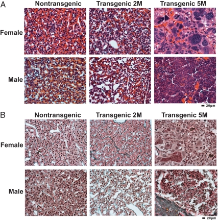
Pituitary histology of GnRHR-TAg transgenic mice reveals a sex-specific difference in tumor severity. A, H&E staining of female and male pituitary tissues from nontransgenic and GnRHR-TAg transgenic mice at 2 months of age (2M) revealed normal histology in both sexes. At 5 months of age (5M), both transgenic females and males displayed transformed cellular histology, but distinct in severity. B, Histological examination with reticulin staining of same tissue in A, demonstrating normal tissue organization in 2-month-old female and male nontransgenic and transgenic pituitaries by the presence of reticulin fibers (gray-black). Disruption of reticulin staining indicated tumor formation in transgenic 5-month-old female and male pituitaries with evidence of cellular transformation consistent with pituitary carcinoma in the female pituitary tissue and with that of adenoma in males. Magnification, ×400. Bars, 20 μm.

