Figure 2.
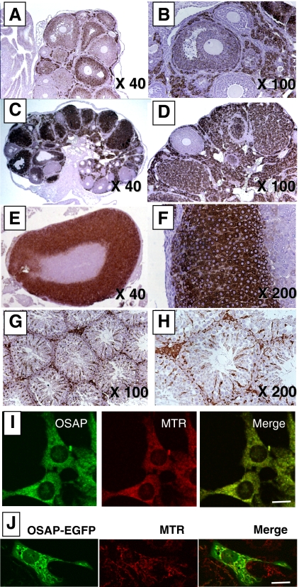
Cellular and subcellular localization of OSAP protein in mice. Immunohistochemical studies were carried out as described in Materials and Methods using ovaries collected 48 h after the administration of PMSG (A and B); ovaries collected 48 h after the administration of hCG to PMSG-primed mice (C and D); adrenal gland (E and F); and testis (G and H). Anti-OSAP antibody immunostaining (left panel) and Mitotracker Red CMX Ros (MTR; a mitochondrial marker) fluorescence (middle panel) show colocalization in Y-1 cells when merged (right panel) (I). Green fluorescence (left panel) generated by a OSAP-EGFP-transfected mouse NIH/3T3 cell and MitoTracker red (MTR; a mitochondrial marker) fluorescence (middle panel) show colocalization when merged (right panel) (J). Magnification bar, 10 μm.

