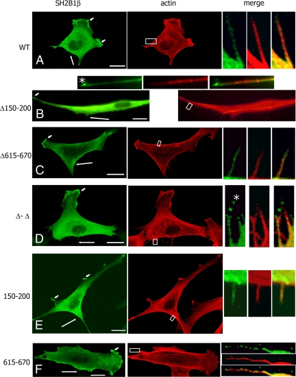
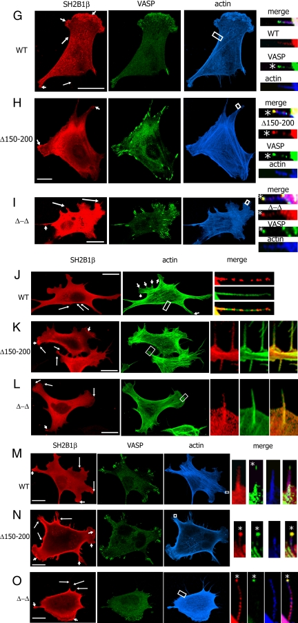
Figure 4.
Intracellular localization of SH2B1β depends on both the first actin-binding domain of SH2B1β and VASP protein. A–F, 3T3 F442A cells overexpressing the indicated forms of myc-SH2B1β were stained with α-myc (green) and phalloidin-rhodamine (red); G–I, 3T3 F442A cells overexpressing GFP-VASP (green) and the indicated forms of myc-SH2B1β were stained with α-myc (red) and phalloidin-647 (blue); J–L, MVD7−/− cells overexpressing the indicated forms of myc-SH21Bβ were stained with α-myc (red), and phalloidin-488 (green). (M-O) MVD7−/− stably overexpressing GFP-VASP (green) were transfected with cDNA encoding the indicated forms of myc-SH2B1β, stained with α-myc (red) and phalloidin-647 (blue). Long arrows indicate filopodia, and short arrows indicate ruffles. The boxed regions were enlarged and merged. Asterisks denote the tips of filopodia. Scale bars, 20 μm.


