Figure 4.
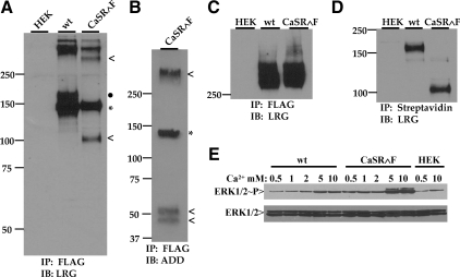
Comparison of wt CaSR and CaSR∧F structure, trafficking, and function. A, Cells expressing CaSR or CaSR∧F were immunoprecipitated with anti-FLAG antibody and Western blotted with anti-CaSR LRG antibody (against residues 374-391 of human CaSR) under reducing conditions (0.1 m DTT) as described in Materials and Methods. Monomeric CaSR forms resolve at 140 kDa (immature, light asterisk) and 160 kDa (mature, filled circle). The immature uncleaved CaSR∧F is observed at 140 kDa (light asterisk), and the larger fragment of the mature, cleaved form (containing LRG epitope) is resolved at approximately 108 kDa (indicated with <). Reduction of receptors is not complete even in 0.1 m DTT, and the cleaved form of CaSR∧F is also apparent in the dimer band region of the blot (indicated with <). B, Western blot of CaSR∧F processed as described in panel A. The blot was probed with anti-CaSR ADD antibody (against residues 214-235 of human CaSR). The immature, uncleaved form (light asterisk) is observed at 140 kDa, and the cleaved fragments containing the ADD epitope are resolved as a doublet at 48/54 kDa (indicated with <). C, Cells transfected with either CaSR or CaSR∧F immunoprecipitated with anti-FLAG antibody in the absence of reducing agents were run on Western blots and probed with anti-CaSR LRG antibody. Both wt and CaSR∧F are resolved in dimeric form above 250 kDa. D, Cells expressing wt or CaSR∧F were biotinylated as described in Materials and Methods and proteins isolated with streptavidin-agarose. Samples were eluted under reducing conditions and Western blots probed with anti-CaSR LRG antibody. E, Transiently transfected cells were starved in 0.5 mm Ca2+/0.2% BSA in DMEM for 12 h before stimulation with varying extracellular Ca2+ for 10 min 37 C. Lysates were processed for Western blotting with anti-phospho-ERK1/2 antibody, stripped, and reprobed with anti-ERK1/2 antibody. Untransfected HEK293 cells stimulated with either 0.5 or 10 mm Ca2+ were used as controls. IB, Immunoblotting; IP, immunoprecipitation.

