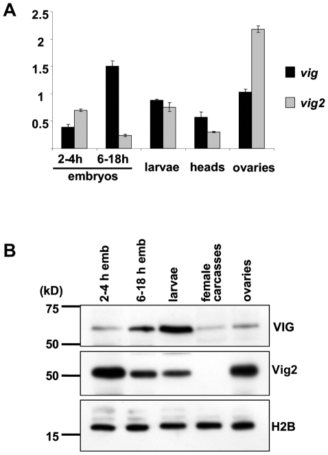
Figure 1. A developmental profile of expression of the vig and vig2 genes generated using quantitative real-time RT PCR and Western immunoblotting.
(A) Vig and vig2 are transcriptionally active throughout development reaching the highest levels in ovaries (vig2) and in larvae (vig). Vig and CG11844 expression levels shown are normalized to the expression of the RpL32 gene. (B) VIG protein can be detected at all stages of development and in somatic as well as in germline tissue (Western blot using CSH1801). Significant protein accumulation occurs in late embryogenesis and at the larval stage. Vig2 protein is at its highest levels in ovarian tissue and early embryos, but is present throughout development; the amount of protein gradually declines and becomes undetectable in adult soma (Western blot using CSH2542). H2B antibodies were used for the loading control.

