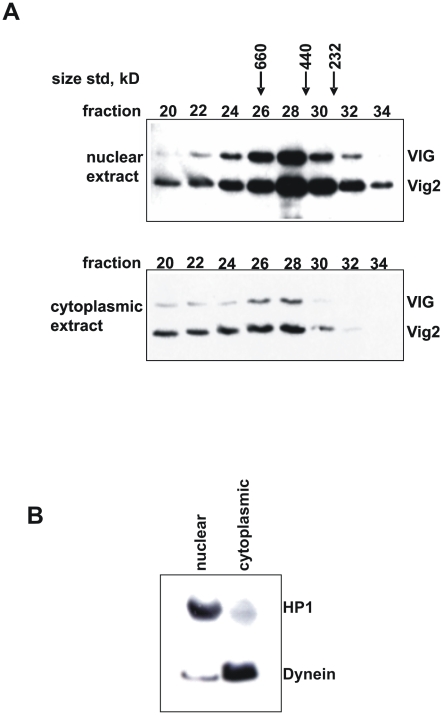
Figure 2. Recovery of VIG and Vig2 from nuclear and cytoplasmic S2 cell extracts.
(A) Nuclear and cytoplasmic material was subjected to Superose-6 column chromatography. The numbers above the Western image represent different fractions. Peaks of size standards used to calibrate the column are shown. The Western blot was performed using an antiserum that recognizes both VIG1 and Vig2 (CSH1803). (B) Segregation of nuclear and cytoplasmic components in the extracts used for column chromatography is verified by a control Western blot for HP1 (nuclear) and Dynein (cytoplasmic).

