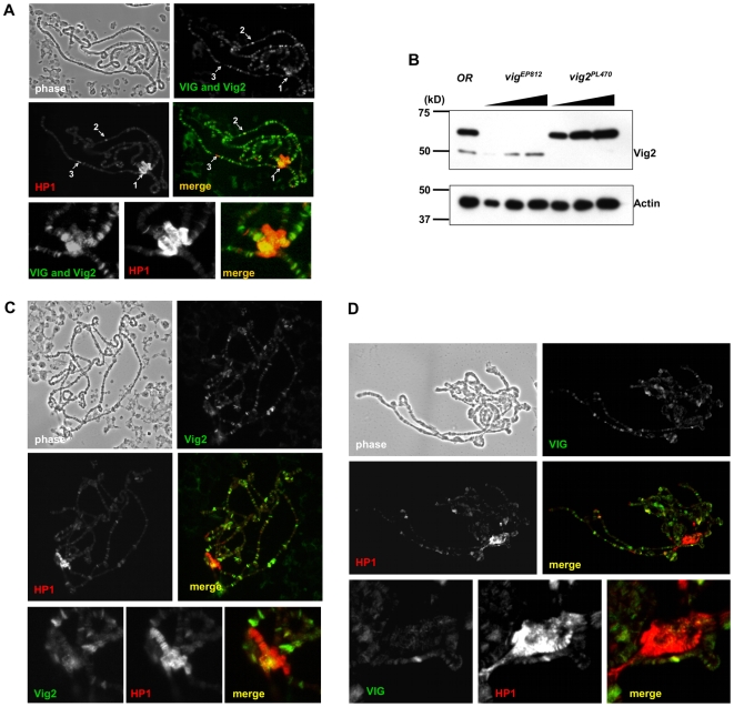
Figure 4. Immunofluorescent staining of polytene chromosomes.
(A) VIG and Vig2 (visualized using CSH1803) show a distribution pattern that overlaps with HP1 at the chromocenter (arrow 1) and in some cases on the chromosome arms (arrow 2) of the WT polytene chromosomes. However, the majority of the cytobands do not overlap; for instance, region 31 on the 2nd chromosome (the ‘goose neck’) is associated with HP1 alone (arrow 3). The lower row of images shows staining at the chromocenter. (B) Western blot performed using protein from salivary glands shows detection of both VIG and Vig2 protein by the CSH1803 antibody. In the vigEP812 mutant only Vig2 is present; in the cg11844PL470 mutant only VIG is recognized. (C) Polytene chromosomes were obtained from vigEP812 mutant larvae; in the absence of VIG we observe significant overlap between HP1 and Vig2 protein at the chromocenter and at some sites on the chromosome arms. The lower row of images shows staining at the chromocenter. (D) The distribution of VIG seen on vig2PL470 mutant polytene chromosomes shows multiple overlapping sites shared by HP1 and VIG on the chromosomal arms, but only faint speckles of VIG at the chromocenter. The lower row of images shows staining at the chromocenter.

