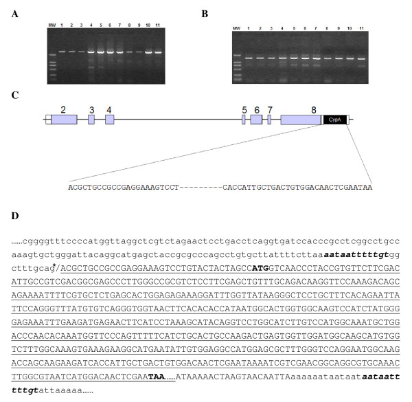
Figure 1.
Formation of TRIMCyp fusion gene in M. leonina. (A and B) The genomic sequences spanning from the 5' end of exon 8 to the 3' end of exon 8 (A) or to the 3'-UTR of CypA cDNA (B) were PCR amplified and subject to electrophoresis analysis. MW: DNA molecular weight marker DL-2000; Lane 1–11: M. leonina samples 524, 528, 551, KMZ-1, KMZ-2, KMZ-4, KMZ-5, 87015, 93201, 97203, and 99201. (C) Schematic structure of the TRIM5Cyp fusion. The exons are represented as boxes with the coding region being shaded, and the sequence of the inserted CypA cDNA is denoted below. (D) The CypA pseudogene cDNA retrotransposed into TRIM5 locus. The asterisk (*) indicates the splicing acceptor, CypA pseudogene cDNA sequence is underlined, target site duplication (TSD) is in bold italic, and the start or stop codon of inserted CypA cDNA is in bold-type.

