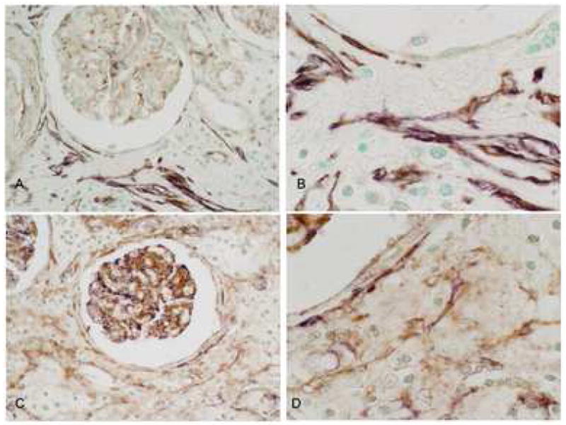
Fig. 4.

PDGF-D colocalization with smooth muscle actin and PDGF-Rβ in the interstitium. A and B: Double IHC showing PDGF-D (brown) and α-smooth muscle actin (purple). PDGF-D is expressed by glomerular podocytes and a subset of interstitial cells (A). High power view demonstrates colocalization of PDGF-D and α-smooth muscle actin by interstitial myofibroblasts.
C and D. Double IHC showing PDGF-D (purple) and PDGFR-beta (brown). PDGF-D is expressed by glomerular podocytes and a subset of interstitial cells (C). High power view shows PDGF-D colocalized PDGFR-beta in a subset of interstitial myofibroblasts (D). Original magnification A and C ×400, B and D ×1000.
