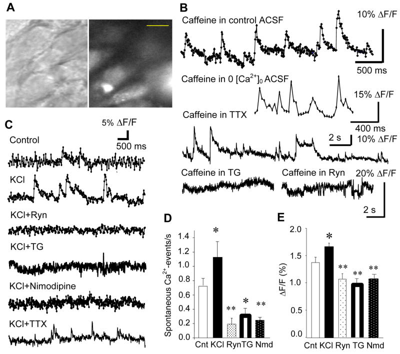
Figure 1.
Miniature [Ca2+]i events in the CA1 pyramidal cell. A. Visually-identified CA1 pyramidal cells with DIC (left) were loaded with fluo-3AM (right). Calibration bar: 10 μm. B. Caffeine enhanced the probability of generating miniature [Ca2+]i-events, which persisted in the Ca2+-free ACSF and in the tetrodotoxin (TTX, 1 μM)-containing ACSF. However, caffeine was not effective of generating any [Ca2+]i-event in the presence of thapsigargin (TG, 4 μM) and ryanodine (Ryn, 10 μM). C. Miniature [Ca2+]i-events in the control ACSF, 80 mM K+-containing ACSF, 80 mM K+ and ryanodine-containing ACSF, 80 mM K+ and TG-containing ACSF, 80 mM K+ and nimodipine-containing ACSF, and 80 mM K+ and TTX-containing ACSF. D. Mean frequency of miniature [Ca2+]i-events in the above 5 experimental conditions (excluding TTX experiment, see text for explanations). E. Mean amplitude (measured as %ΔF/F0) of miniature [Ca2+]i events in the 5 conditions. Cnt: Control, KCl: 80mM K+-containing ACSF, Ryn: ryanodine and 80 mM K+-containing ACSF, TG: thapsigargin and 80 mM K+-containing ACSF, Nmd: nimodipine and 80 mM K+-containing ACSF.

