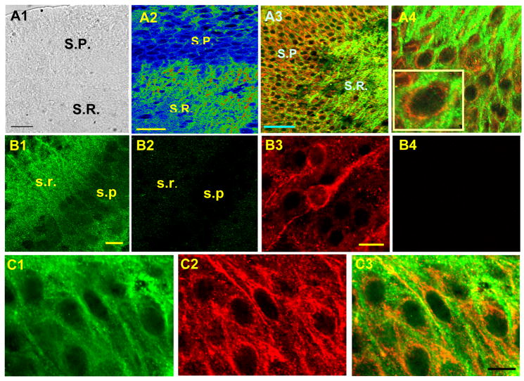
Figure 3.
Juxtaposed localization of RyR and LTCC (CaV1.2). A1. A bright field image of the hippocampal slice culture that was used for dual labeling of RyR and CaV1.2. A2. CaV1.2 immunoreactivity in CA1 (warmer color represents stronger immunoreactivity). A3. Dual labeling of RyR (red) and CaV1.2 (green). SP: Stratum Pyramidale, SR: Stratum Radiatum. A4. Dual labeling in the pyramidal cell layer with higher magnification. B1. CaV1.2. B2. CaV1.2 with a blocking peptide. B3. RyR, B4. RyR with a blocking peptide. C1–3. Dual labeling of CaV1.2 and RyR with an image of CaV1.2 only (C1), RyR only (C2), and a merged image of both CaV1.3 and RyR (C3). These three images were taken sequentially from the identical population of the neurons in the same slice. Calibration bar: 50 μm in A1 and A2, 20 μm in B1 and B3, 10 μm in C3.

