Fig. 2.
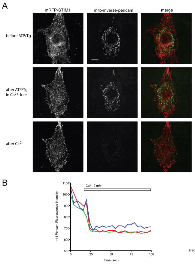
Confocal image of a COS-7 cell expressing mRFP-STIM1, untagged Orai1 and inverse-Pericam-targeted to the mitochondrial matrix. (A) Upper row: control, middle row: 2 min after stimulation with 50 μM ATP and 200 nM Tg in Ca2+-free medium (note the formation of STIM1 puncta on the left panels), lower row: 2 min after the addition of 2 mM Ca2+ (note the disappearance of i-Pericam fluorescence). The bar shows 10 μm (B) The lower panel shows the rapid fall of mitochondrially targeted i-Pericam (i.e. the increase in [Ca2+]m) in 3 separate mitochondrial ROIs in response to readdition of Ca2+. (Performed at 35 C.)

