Figure 6.
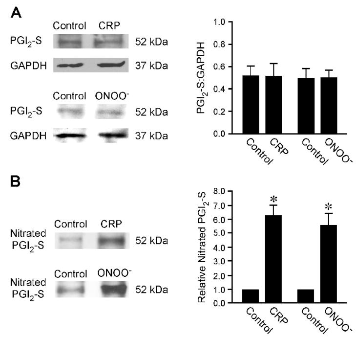
Expression of prostacyclin synthase (PGI2-S) and nitrated PGI2-S in isolated coronary arterioles. (A) The left panel shows immunoblots of PGI2-S and GAPDH in vessels treated with vehicle (Control), CRP (7 μg/mL), or peroxynitrite (ONOO−, 10 μM, positive control) for 60 minutes. The right panel shows the total PGI2-S protein normalized with corresponding GAPDH protein. CRP or peroxynitrite treatments did not change the total PGI2-S expression in coronary arterioles. Data represent three independent experiments. (B) The left panel shows immunoblots of nitrated PGI2-S in nitrotyrosine immunoprecipitates from vessels treated with vehicle (Control), CRP (7 μg/mL), or peroxynitrite (ONOO−, 10 μM) for 60 minutes. The right panel shows the relative amount of nitrated PGI2-S. The amount of nitrated PGI2-S was significantly increased in CRP- and peroxynitrite-treated coronary arterioles. Data represent three independent experiments. *P < 0.05 vs. Control.

