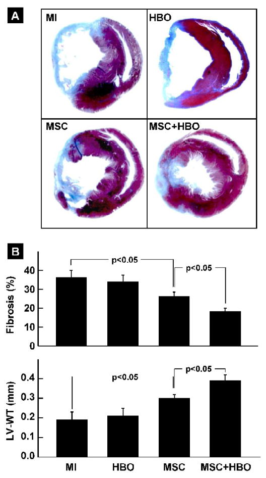
Figure 5. Cardiac tissue fibrosis and remodeling in MI hearts treated with MSC and HBO.
Masson-trichrome staining of heart sections was performed in infarcted hearts (MI), and infarcted hearts treated with HBO (HBO), MSC (MSC) or MSC followed by HBO (MSC+HBO) at 2 weeks after transplantation of MSCs. (A) Representative images of heart sections stained with Masson-trichrome. (B) Percentage of fibrosis and LV wall thickness (LV-WT), as determined by computer planimetry. Data represent mean±SD obtained from 6 hearts per group. The MSC+HBO group exhibits a significant reduction in the fibrosis and improvement in LV-WT when compared to MSC group.

