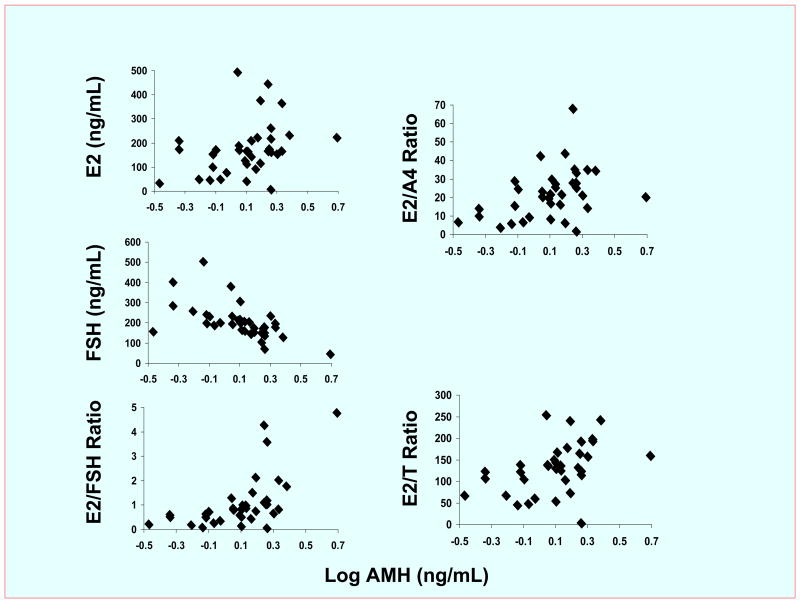
Figure 1.
Correlations between AMH and hormone concentrations in follicles of 26 normoandrogenic ovulatory women undergoing IVF-ET. AMH levels significantly correlated with E2 (R=+0.26; P<0.025) and FSH (R=−0.74; P<0.0001) concentrations as well as E2/FSH (R=+0.70; P<0.000), E2/A4 (R=+0.26; P<0.025) and E2/T (R=+0.31; P<0.0005) ratios. Conversion to SI units, AMH* 7.14 pmol/L. The first follicle of each ovary was selected by size (at least 16 mm in diameter) and accessibility to replicate the follicle size in which AMH previously has been shown to predict enhanced oocyte development (6).

