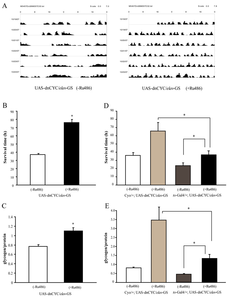
Figure 7. Clocks in neuronal cells oppose effects of the fat body clock on starvation and energy stores.
(A) Expression of dnCYC in neuronal cells abolishes the locomotor activity rhythm. Adult male flies expressing dnCYC in neuronal cells display arrhythmic locomotor activity in DD. (B) Flies expressing dnCYC in neuronal cells are less sensitive to starvation than control male flies. Asterisks indicate significant differences (p<0.05). (C) Glycogen storage is significantly higher in flies expressing dnCYC in neuronal cells as compared to control male flies. (D) and (E) Flies with both neuronal and fat body clocks disrupted have a normal response to starvation and normal glycogen stores. Flies carrying UAS-dnCYC and elav-GS-Gal4 with or without to-Gal4 were treated with RU486 to induce expression of elav-GS. In the absence of RU486, to-Gal4/+;UAS-dnCYC/elav-GS flies are more sensitive to starvation (D) and have less glycogen (E) than controls that lack to-Gal4. This is because disruption of the fat body clock increases sensitivity to starvation. However, after treatment with RU486 to induce additional disruption of neuronal clocks, the starvation response and glycogen levels are similar to those in wild type. Note that for the experiments in Figure 7D and 7E flies were treated with RU484 for ~2 weeks as compared to a 5–6 day treatment in Figure 7A –7C; this most likely accounts for the difference in the glycogen increase. Asterisks indicate significant differences (p<0.05). Statistical significance was determined by two-tailed Student’s t-test with unequal variance.

