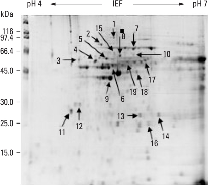
Fig. 2.
Proteome pattern of cervical cancer tissue. Nineteen protein spots on the gel were marked with arrows. Numbered spots were excised from the cancer tissue gel, in-gel digested with trypsin, and identified by MALDI-TOF assay. The results are listed in Table 2. MALDI-TOF, matrix assisted laser desorption ionization-Time of flight mass spectrometry.

