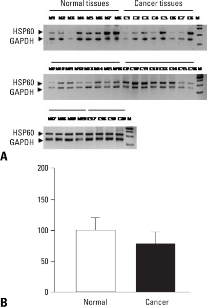
Fig. 3.
(A) RT-PCR analysis of HSP60 mRNA in normal (lane 1-8) and cervical cancer (lane 9-16) tissues. (B) RT-PCR was performed using 1 µg of total RNA and separated on 2.5% agarose gel. The size of PCR products was 320 base pairs. Glyceraldehyde-3-phosphate dehydrogenase (GAPDH) was used as an internal control to confirm equal loading of the samples. HSP60 mRNA levels were quantified as a percentage of relative optical density. Results are mean ± S.E.M. of 20 samples per group. RT-PCR, reverse transcriptase polymerase chain reaction; mRNA, messenger RNA; HSP60, heat shock protein.

