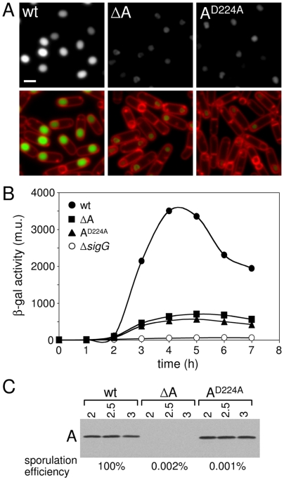
Figure 1. The ATPase motifs in SpoIIIAA are required for σG activity and sporulation efficiency.
(A) σG activity was assessed in single cells by microscopy using a fluorescent reporter (PsspE-cfp) in a wild-type background (wt, BTD2779), a ΔspoIIIAA mutant (ΔA, BTD2713) and a spoIIIAA D224A mutant (AD224A, BTD2775). Cells were visualized at hour 3 of sporulation. Forespore CFP fluorescence (false-colored green in the lower panel) and the fluorescent membrane dye TMA-DPH (false-colored red) are shown. Scale bar, 1 µm. (B) Expression of a σG-dependent sspB-lacZ translational fusion [53] was monitored during a time course of sporulation in a wild-type background (wt, BTD2919), a ΔspoIIIAA mutant (ΔA, BTD2917), a spoIIIAA D224A mutant (AD224A, BTD2920) and a strain lacking σG (ΔsigG, BTD1331). Samples from sporulating cells were taken every hour and β-galactosidase activity (Miller Unit, m.u.) was determined. (C) Immunoblot analysis of whole cell lysates from sporulating cells shown in A using anti-SpoIIIAA antibodies. Time (in hours) after the initiation of sporulation is indicated. Sporulation efficiencies of the same strains are shown below the immunoblot.

