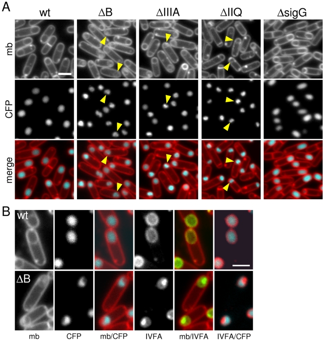
Figure 4. Morphological defects in the absence of SpoIIIA or SpoIIQ.
(A) Forespore morphology was monitored by fluorescence microscopy at hour 3 of sporulation in a wild-type background (wt, BCM703), a ΔspoIIIAB mutant (ΔB, BCM704), a ΔspoIIIA mutant (ΔIIIA, BCM706), a ΔspoIIQ mutant (ΔIIQ, BCM716), and in a strain lacking σG (ΔsigG, BCM708). All strains contained a forespore reporter (PspoIIQ-cfp; false-colored blue in the lower panel) to visualize the forespore cytoplasm. The membranes (mb) from the same field were visualized with the fluorescent dye TMA-DPH (false-colored red in the lower panel). Carets highlight examples of “collapsed” forespores. (B) Larger images highlighting the cytoplasmic CFP signal and the localization YFP-SpoIVFA (IVFA; false-colored green) in wild-type (wt, BCM703) and a ΔspoIIIAB mutant (ΔB, BCM704). Larger images of all strains can be found in Figure S5. Scale bar, 1 µm.

