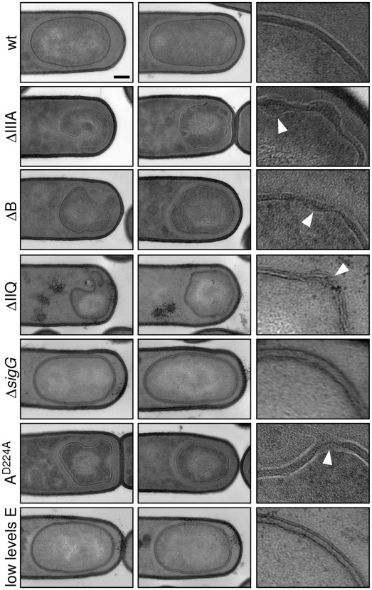
Figure 5. Morphological defects in the absence of the SpoIIIA or SpoIIQ proteins.
Forespore morphology was assessed by electron microscopy at hour 3 of sporulation in wild-type (wt, PY79), a ΔspoIIIA mutant (ΔIIIA, BDR841), a ΔspoIIIAB mutant (ΔB, BTD119), a ΔspoIIQ mutant (ΔIIQ, BTD141), a ΔsigG mutant (ΔsigG, BDR104), a spoIIIAA D224A ATPase mutant (AD224A, BTD2683), and a strain that contains low levels of SpoIIIAE (low E, BTD3019). For each strain, two typical forespores are shown in the first two columns. Scale bar, 200 nm. The last column shows a 5× enlargement of the forespore membranes. The carets highlight bulged or ruptured membranes in the mutants.

