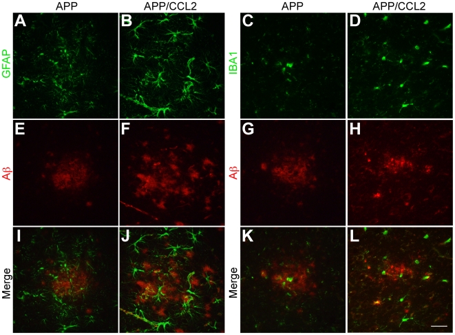
Figure 4. Accumulation of astrocytes and microglia to Aβ deposits.
Frozen sections (10-µm thickness) of temporal cortex of APP and APP/CCL2 mice at 9 months of age were subjected to double immunofluorescence for GFAP (A–B, green) or IBA1 (C–D, green) and Aβ (NU-1) (E-H, red). (I–L) Merged images. Scale bar, 50 µm.

