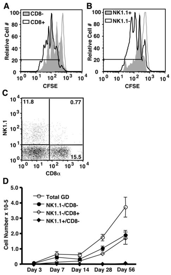
Figure 1. CD8+ and NK1.1+ γδ T cell subsets undergo homeostatic expansion in TCRβ−/−δ−/− mice at distinct rates.
(A and B) Splenic γδ T cells were isolated from TCRβ−/− mice, labeled with CFSE, and injected into TCRβ−/−/δ−/− recipients. Homeostatic proliferation of CD8− and CD8+ or NK1.1− and NK1.1+ γδ T cells subsets was assessed on day 5 by flow cytometry. Gates delineate those cells that have undergone at least one division, as determined by the CFSE levels of non-T cells within our donor population (unfilled grey histogram). (C) The frequency of NK1.1−/CD8−, NK1.1−/CD8+, and NK1.1+/CD8− in the donor γδ T cell population was determined by flow cytometry. Percentages of live CD3+/TCRδ+ cells are shown for each quadrant. (D) γδ T cell reconstitution was assessed from 3 days to 2 months after transfer into TCRβ−/−/δ−/− mice.

