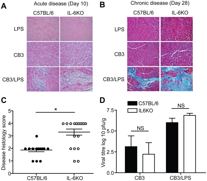
Figure 1. IL-6KO mice develop increased chronic myocarditis severity without an increase in virus in the heart.
(A) Representative Hematoxylin and Eosin stained cardiac sections from C57BL/6 or IL-6KO mice. Mice treated with LPS or CB3 alone did not develop acute disease lesions at day 10 PI (LPS: C57BL/6 n = 4, IL-6KO n = 4) (CB3: C57BL/6 n = 5, IL-6KO n = 4). Mice treated with CB3/LPS developed lesions and immune cell infiltration at 10 days PI (C57BL/6 n = 4, IL-6KO n = 4). Magnification:400×. (B) Representative Masson's Trichrome stained cardiac section from C57BL/6 or IL-6KO mice. Following treatment with CB3/LPS, mice developed disease pathology as determined by fibrosis in blue and immune cell infiltration within the fibrosis areas by 28 days PI (C57BL/6 n = 15, IL-6KO n = 20). Mice infected with LPS or CB3 alone did not develop significant chronic disease pathology (LPS: C57BL/6 n = 5, IL-6KO n = 5) (CB3: C57BL/6 n = 6, IL-6KO n = 7). Magnification:400×. (C) Chronic cardiac disease histology was scored blindly by a four tier grading system to determine severity differences: grade 1, 0–10% pathology; grade 2, 11–25%; grade 3, 26–50%; grade 4, greater than 50% (black circles indicate wt mice, white circles indicate IL-6KO mice) (bar is mean±SE, *p<0.05). Disease severity was found to be significantly higher in the IL-6KO mice compared to wild type controls. (D) Quantification of replicative virus in the heart post infection showed no significant differences in viral titer between IL-6KO and wt mice at day 5 post infection (CB3: C57BL/6 n = 5, IL-6KO n = 5) (CB3/LPS: C57BL/6 n = 5, IL-6KO n = 5) (black bars indicate wt mice, white bars indicate IL-6KO mice) (mean±SE, NS = not significant). This indicates that IL-6 is not essential for control of viral replication in the heart.

