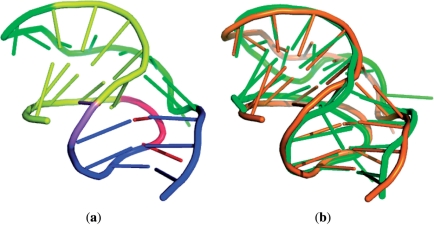
Figure 6.
(a) Tertiary structure of a frameshifting pseudoknot (PDB ID: 1YG3, chain: A, nucleotide numbers: 3–30). Stem 1 is in yellow, stem 2 is in blue, loop 1 is in red, loop 2 is in green and the nucleotide (A13) between the two stems is in violet. (b) The superposition between the query pseudoknot (1YG3) colored orange and an identified pseudoknot (2AP5) colored green with an RMSD of 2.97 Å.

