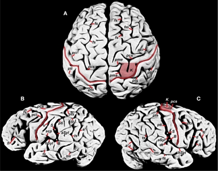
Figure 1.
Photographs of Einstein's brain that were taken in 1955, adapted from Witelson et al. (1999b) with identifications added here. (A) Dorsal view, (B) left lateral view, (C) right lateral view. Sulci: angular (a2), anterior occipital (a3), ascending limb of the posterior Sylvian fissure (aSyl), central fissure (red lines), diagonal (d), descending terminal portion of aSyl (dt), inferior frontal (fi), middle frontal (fm), superior frontal (fs), horizontal limb of the posterior Sylvian fissure (hSyl), intraparietal (ip), precentral inferior and superior (pci, pcs), marginal precentral (pma), medial precentral (pme), postcentral inferior and superior (pti, pts), ascending ramus of Sylvian fissure (R), subcentral posterior sulcus (scp), middle temporal (tm), superior temporal sulcus (ts), unnamed sulcus in postcentral gyrus (u). Other features: branching point between hSyl and aSyl (white dots, B), hand motor cortex knob (K, shaded in A, C), termination of aSyl (white dots, S).

