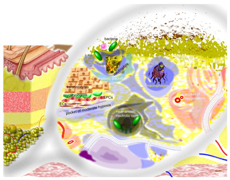
Figure 2. Heterogeneous distribution of oxygen in the wound tissue: hypothetical pockets of graded levels of hypoxia.
Structures outside the illustrated magnifying glass represent the macro tissue structures. Objects under the glass represent a higher resolution. Shade of black (anoxia) or blue represents graded hypoxia. Shade of red or pink represents oxygenated tissue. Tissue around each blood vessel is dark pink in shade representing regions that are well oxygenated (oxygen-rich pockets). Bacteria and bacterial infection are presented by shades of green on the surface of the open wound.

