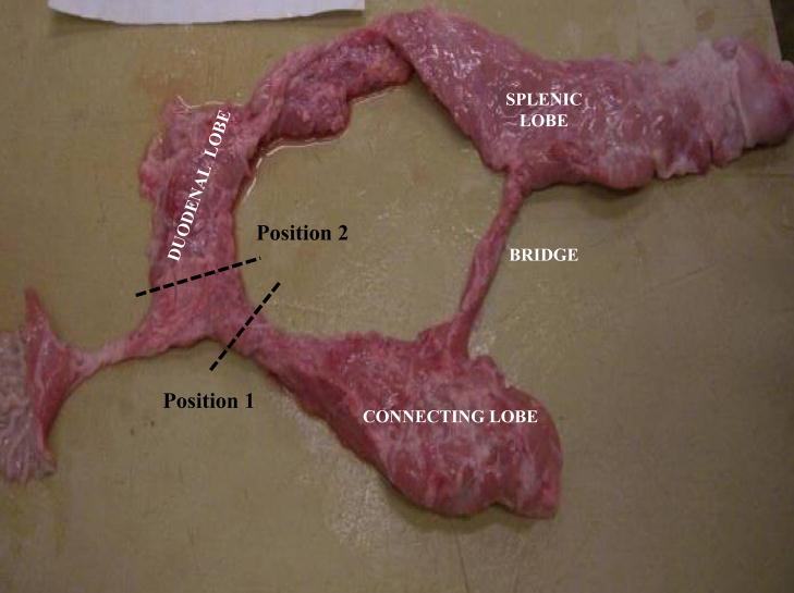
Figure 1.
Photograph of an excised pig pancreas exhibiting normal anatomy with the duodenal, splenic, and connecting lobes, as well as the bridge.
Position 1: Dotted lines indicate the positioning of the clamp restricting flow to the connecting lobe.
Position 2: Dotted lines indicate the positioning of the clamp restricting flow to the duodenal lobe.

