
FIGURE 9. Two-dimensional representation of a proposed three-dimensional model illustrating the synthetic melanocortin NDP-MSH docked inside the hMC4R.
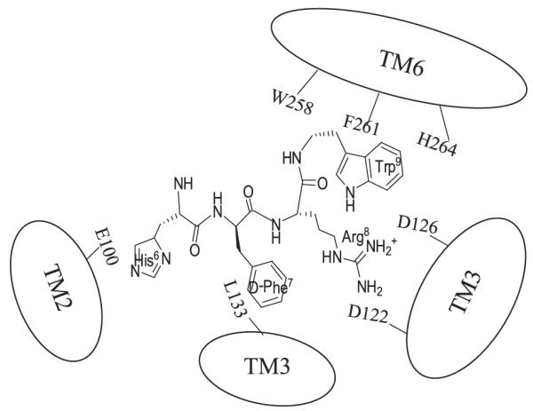
Two receptor binding pockets are hypothesized. The first is a predominantly ionic pocket formed by Glu100, Asp122, and Asp126. The second hydrophobic pocket is formed by aromatic residues Trp258, Phe261, and His264 in TM6.