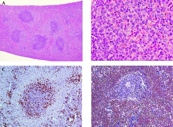
Figure 4.
Representative histology and immunohistochemistry of splenic marginal zone lymphomas that do not express CD45R/B220. (A) Splenic marginal zone lymphoma with multiple follicles that have expanding marginal zones that extend into the red pulp and bridge the follicles. Hematoxylin and eosin stain; magnification, ×4. (B) Splenic marginal zone lymphoma in the red pulp. The lymphoma cells have small to medium round or irregular nuclei and abundant pale cytoplasm. A mitotic figure is evident in the upper right corner. Hematoxylin and eosin stain; magnification, ×50. (C) The lymphoma cells in the marginal zone surrounding the follicle and in the red pulp do not express CD45R/B220. Magnification, ×10. (D) The lymphoma cells in the marginal zone surrounding the follicle and filling the red pulp express Pax5 with strong intensity. Magnification, ×10.

